Light in the black box
Freiburg, Nov 10, 2017
The brain is a black box. Understanding it takes more than opening the cover and looking inside. It's too complex. Neurophysiology has long been trying to discover more – by measuring cell activity with electrodes, for example. The aim is to find out what happens in the brain during particular movements. Biology professor llka Diester, who is also a member of the BrainLinks-BrainTools excellence cluster, has a completely different approach to the brain. "We activate cells in a spatially and temporally selective way, and then look at what happens," she explains. Diester adds that's the only way to find out if specific cells are relevant to a precise behavior. The 39-year-old has received a European Research Council (ERC) Starting Grant for her work. The University of Freiburg is celebrating the tenth anniversary of the ERC Grant and its 50 award-winners with a look at selected projects. A series introduces ten prize-winning researchers and their work.
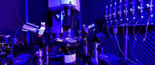
 Hightech in neurological research: The team works with photon microscopes to find out which cells are responsible for which behaviors.
Photo: Thomas Kunz
The focus of Ilka Diester's research is the brain's motor cortex, which is involved in voluntary movements. "We want to know how it communicates with the adjacent areas and what kind of behavioral output this communication has," says Diester. She adds that the 1.5 million euro ERC Grant provides tremendous support. The funding enabled her to get expensive devices and employ doctoral candidates. The team is using something called optogenetic technology in their work. It's supposed to shine a bit of light into the black box of the brain. Literally. The technology is based on membrane proteins that are sensitive to light in a way similar to proteins found in green algae. After finishing her biology degree and doctorate, Diester went to Stanford in the US to do postdoctoral work for three years under, among others, Karl Deisseroth, whom she calls the "godfather" of optogenetics. That is where Diester became familiar with the technology.
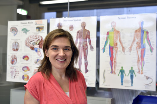
 Ilka Diester wants to find out what happens in the brain during specific movements.
Photo: Thomas Kunz
But let's get back to green algae, which requires opsins as well as light for photosynthesis. Opsins are light sensitive molecules that are found in the membrane proteins of the cell envelope. They function as guides, showing the algae exactly where the light source is. Diester gets specific, "When light in the form of a photon hits the opsin, it opens and lets ions flow through the membrane. Positively charged ions, for example. These increase the membrane potential which opens additional voltage-dependent channels." And the green algae begins to move. Says Diester, "It becomes active, and shifts from a lazy, paddling rhythm into the beat of a powerful breast stroke and moves towards the light."
Genetic tricks
But opsins are only found in microorganisms, not mammals. Still, with a bit of genetic slight-of-hand, the opsins can be introduced into mammalian neurons, making them light sensitive. A DNA structure made up of three components – the opsin gene, the promoter and a fluorescence protein gene – is placed in the cell. "You can imagine it as a kind of shuttle service," explains Diester, "the DNA is placed in a weakened virus – the shuttle – and injected into the mouse's brain." There, the opsin is deposited in the neural membrane. The promoter determines the cells in which the opsin gene is expressed as opsin protein, while the fluorescence gene marks these cells.
"That way, we can monitor the neurons in time and space," explains Diester. Or, to be more precise, disturb them. The stimulation of the lab animal's cells is done locally. It has a small implant in its head to which a fiber-optic cable can be connected. Right alongside it is the tip of a tiny electrode that measures exactly what happens. As soon as light hits the opsins, the nerve cells are activated or deactivated.
Manipulate and observe
The cells keep functioning normally in spite of the opsin that's been introduced. Yet manipulation takes place during exposure to light and the researchers observe. What does the mouse do if the researchers inhibit these cells? Does the mouse still find it's way to food? Or does it keep going around in circles? Action potentials move from neuron to neuron, networking different areas of the brain. Intentionally stimulating or inhibiting select communication pathways between different areas of the brain disrupts neural interaction. This allows researchers to determine how relevant individual pathways are for behavior. That's what the ERC project is all about.
Understanding how the motor cortex communicates with other areas of the brain could help in treating humans with impulse control disorders. That's the long-term goal, says Diester, adding, "opotogenetics may seem far-out, but it's not that inconceivable."
Stephanie Streif

